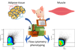


The adaptability of livestock to changing environments is based in particular on their genetic characteristics but also on the farming conditions to which they are subjected. However, this last point is poorly documented and little is known about its contribution to environmental challenges. The study by Quéméner and colleagues [1] addresses this question by assessing the effect of two hygiene conditions (good vs poor) on the distribution of cell populations present in adipose and muscle tissues of pigs divergently selected for feed efficiency [2].
The working hypothesis is that degraded housing conditions would be at the origin of an hyper stimulation of the immune system that can influence the homeostasis of adipose tissue and skeletal muscle and consequently modulate the cellular content of these tissues. Cellular compositions are thus interesting intermediate phenotypes for quantifying complex traits. The study uses pigs divergently selected for residual feed intake (RFI+ and RFI-) to assess whether there is a genetic effect associated with the observed phenotypes.
The study characterized different stromal cell populations based on the expression of surface markers: CD45 to separate hematopoietic lineages and markers associated with the stem properties of mesenchymal cells: CD56, CD34, CD38 and CD140a. The authors observed that certain subpopulations are differentially enriched according to the hygiene condition (good vs poor) in adipose and skeletal tissue (CD45-CD56-) sometimes with an associated (genetic) lineage effect. This pioneering study validates a number of tools for characterizing cell subpopulations present in porcine adipose and muscle tissue. It confirms that housing conditions can have an effect on intermediate phenotypes such as intra-tissue cell populations. This pioneering work will pave the way to better understand the effects of livestock systems on tissue biology and animal phenotypes and to characterize the nature and function of progenitor cells present in muscle and adipose tissue.
[1] Quéméner A, Dessauge F, Perruchot MH, Le Floc’h N, Louveau I. 2022. The impact of housing conditions on porcine mesenchymal stromal/stem cell populations differ between adipose tissue and skeletal muscle. bioRxiv 2021.06.08.447546, ver. 3 peer-reviewed and recommended by Peer Community in Animal Science. https://doi.org/10.1101/2021.06.08.447546
[2] Gilbert H, Bidanel J-P, Gruand J, Caritez J-C, Billon Y, Guillouet P, Lagant H, Noblet J, Sellier P. 2007. Genetic parameters for residual feed intake in growing pigs, with emphasis on genetic relationships with carcass and meat quality traits. Journal of Animal Science 85:3182–3188. https://doi.org/10.2527/jas.2006-590.
DOI or URL of the preprint: https://doi.org/10.1101/2021.06.08.447546
Dear Editor,
All listed authors, and I as corresponding author, are very grateful for your consideration about our manuscript. We are pleased to submit a copy of our revised manuscript entitled "The impact of housing conditions on porcine mesenchymal stromal/stem cell populations differ between adipose tissue and skeletal muscle", which has been modified according to appropriate and helpful comments of the two reviewers.
We hope that the revised manuscript reaches the standards required for publication in PCI Animal Science.
Sincerely yours,
Isabelle Louveau
INRAE
UMR1348 Pegase
F-35590 Saint Gilles
France
E-mail : isabelle.louveau@inrae.fr
Dear Dr Louveau,
Your preprint entitled " The impact of housing conditions on porcine adult stem cell populations differ between adipose tissue and skeletal muscle" has now been seen by 2 referees. You will see from their comments below that while they find your work of interest, some important points are raised. We are interested in the possibility of recommending your study in PCI Animal Sciences, but would like to consider your response to these concerns.
We therefore invite you to revise your preprint, taking into account the points raised by your reviewers.
Best regards
Hervé Acloque
This article describes the impact of housing conditions (good vs poor) on the proportion of porcine adult stem cell populations both in adipose tissue and skeletal muscle, in two lines selected for their residual feed intake (HRFI vs LRFI). Stem cell populations are deeply analyzed by a combination of 5 different cell markers (CD45, CD34, CD38, CD56 and CD140a) and the results presented here highlight some differences in stem cell populations after the sanitary challenge, with differences observed between adipose tissue and skeletal muscle of both RFI lines.
If the hypotheses behind the search for differences within tissues and within the sanitary challenge are well explained in the introduction, the rationale of the use of the two RFI lines is not obviously stated. What was expected in those two lines? What differences in the responses to the sanitary challenge between the RFI lines published by Chatelet et al, 2018 justify doing this study on both lines? Could also the results of the comparison of the 2 lines be more deeply discussed?
The reference to “the period 1 of a larger study” (L133), is not clear. Housing conditions are per se sufficiently developed in the M&M.
For the flow cytometry analysis, the ratio of dispensed cells/tube vs acquired events seems odd for SCAT (L183-192): 50,000 cells were dispensed for SCAT and a minimum of 50,000 events were acquired. In addition, the viability marker used is not provided.
In Table 1: it would be appreciated if the final dilution/concentration of antibodies used could be mentioned.
The statistical analyses used are two-way ANOVA. The effects per group (5 to 9) would suggest to use non parametric test. Please justify the test used.
In Tables 2, 3 and 4: the exact n per group should be clearly stated.
Figure 1 is confusing and does not fit exactly with the gating strategies proposed in Figures 2 and 3. Wouldn’t it be easier to clearly mention the panel of antibodies used for each tissue?
Gating strategies illustrated in Figures 2 and 3 seem not complete: in Figure 2, CD45-CD56- cells are not gated in red and the expression of CD34 within those cells not shown as it is for the CD45-CD56+ cells, but results of those cell populations are reported in Table 3. In Figure 3, also for CD45-CD56- cells, no further gating is shown for CD34 and CD140a expressions but results for CD45-CD56-CD34+, CD45-CD56-CD34-, CD45-CD56-CD34+CD140a+, CD45-CD56-CD34-CD140a+ are reported in Table 4. In addition, the name of the gate CD45-CD56+CD34+CD140a- is a mistake, CD45-CD56+CD34-CD140a+ cells are shown.
For skeletal muscle cells, the CD45- gate seems very odd. Do the FSC high cells are really CD45+ cells? The viability marker also appears high in those cells and the gating was adapted. Should the gate be adapted as for the viability dye? Otherwise, which hematopoietic cells could it be?
Language remarks:
L50 “compared to” instead of “compared with”
L83 “flow cytometry” instead of “flow-cytometry”
L143 “fed ad libitum with a standard” instead of “fed ad libitum a standard”
L275 “in both SCAT and muscle” instead of “in both at SCAT and muscle”
https://doi.org/10.24072/pci.animsci.100113.rev11https://link.springer.com/article/10.1007/s10561-021-09905-z
This article has a nice image of adipose tissue and the different cells and individual markers. Its just a suggestion.
Download the review https://doi.org/10.24072/pci.animsci.100113.rev12